The apothorax is an anatomical term historically used to describe the upper portion of the human torso, which corresponds closely to what modern medicine calls the thoracic cavity or chest region. This area sits between the neck and abdomen and contains several organs essential for survival, including the heart and lungs. Although the word might sound unfamiliar today, it once appeared in classical anatomical discussions when scholars attempted to categorize the body’s major regions.
Think about the simple act of breathing. When your chest expands and contracts, the movement happens within this region. The apothorax forms a protective chamber surrounded by the rib cage, sternum, and spine, ensuring that the organs responsible for respiration and circulation remain safe from injury.
From a biological perspective, this region acts like the control center of life-sustaining functions. Every heartbeat, every breath, and every movement of oxygen through your bloodstream originates from organs located inside the chest cavity. This is why the apothorax—despite being an older term—remains conceptually important when studying anatomy.
Understanding the apothorax also gives students and medical learners a deeper appreciation of how the human body is organized. The chest isn’t simply an empty space; it’s a sophisticated structure made of bones, muscles, and membranes working together to support breathing, protect organs, and maintain internal pressure.
Etymology and Origin of the Word
The term apothorax originates from ancient Greek roots. The prefix “apo” means “upper,” “away,” or “separate,” while “thorax” refers to the chest or breastplate. When combined, the word roughly describes the upper chest region of the body.
Early anatomists used this term when describing the structural divisions of the torso. Medical knowledge during ancient and medieval periods was still developing, so terminology often varied depending on the author or region. Some scholars used apothorax to highlight the upper section of the thoracic cavity, while others applied it more broadly to the entire chest area.
As anatomy evolved into a modern scientific discipline, standardized terminology became necessary. International anatomical naming systems gradually replaced ambiguous or overlapping words with clearer definitions. Because of this shift, the simpler term “thorax” eventually became the universal label for the chest cavity.
Even though the word apothorax is rarely used in contemporary textbooks, it still appears in historical references and educational discussions. Learning about its origin helps us see how anatomical language evolved and why standardization plays such a crucial role in modern medical communication.
Historical vs Modern Use of the Term Apothorax
Why Apothorax Is Rarely Used Today
Medical terminology has undergone major transformations over the past few centuries. Early anatomical studies often used descriptive language that varied widely between researchers. The term apothorax is one example of a word that gradually disappeared as anatomy became more standardized.
One major reason for its decline is inconsistency in definition. Different sources described the apothorax in slightly different ways—sometimes referring to the upper thorax, sometimes to the area around the thoracic cavity, and occasionally to regions bordering the diaphragm. This lack of precision created confusion for physicians and researchers.
To solve this issue, scientific organizations introduced standardized anatomical terminology systems such as Terminologia Anatomica, which replaced ambiguous terms with universally accepted names. In these systems, the word thorax was chosen as the official term for the chest cavity.
The disappearance of apothorax from everyday medical vocabulary doesn’t mean the concept is irrelevant. Instead, it highlights how science evolves. Terminology changes as knowledge improves, allowing professionals across the world to communicate with clarity and precision.
Modern Equivalent: The Thorax
Today, the region once referred to as the apothorax is universally known as the thorax. The thorax is defined as the part of the body between the neck (cervical region) and the abdomen, enclosed by the rib cage and containing vital organs such as the heart and lungs.
Modern anatomical descriptions focus on the thoracic cavity because it provides a clear and consistent framework for understanding the chest. The thorax includes several subdivisions, such as the mediastinum (where the heart is located) and the pleural cavities that house the lungs.
Using standardized terminology also makes medical research and treatment more efficient. When doctors around the world refer to the thorax, they are describing the exact same region. This shared understanding improves collaboration, education, and patient care.
Location of the Apothorax in the Human Body
Position Between Neck and Abdomen
The apothorax occupies a central position in the human body, forming the upper trunk region between the neck and abdomen. This placement allows it to act as a bridge between multiple body systems, including the respiratory, circulatory, and digestive systems.
Imagine the torso as a multi-level building. The neck would be the top floor, the abdomen the lower level, and the apothorax sits right in the middle—housing the most essential mechanical systems that keep the building functioning.
Air enters the body through the nose or mouth, travels down the trachea, and reaches the lungs located within this region. At the same time, the heart pumps oxygen-rich blood from this chamber to every organ in the body.
Because of this central role, the apothorax is surrounded by protective structures. The rib cage forms a curved shell around the chest, while the sternum at the front and the thoracic vertebrae at the back provide structural support.
Anatomical Boundaries of the Apothorax
The apothorax has clearly defined anatomical boundaries that help maintain the structure of the chest cavity and protect its internal organs.
Upper Boundary
The thoracic inlet forms the upper opening of the chest cavity. This small passage connects the apothorax to the neck and allows important structures such as the trachea, esophagus, nerves, and major blood vessels to pass between regions.
Lower Boundary
The lower boundary is created by the diaphragm, a dome-shaped muscle separating the chest cavity from the abdominal cavity. When the diaphragm contracts, it flattens and increases the volume of the chest cavity, allowing the lungs to expand during inhalation.
Structural Components of the Apothorax
Rib Cage and Intercostal Muscles
The rib cage is the most visible structural feature of the apothorax. It consists of twelve pairs of ribs attached to the spine and connected to the sternum by flexible cartilage. This framework protects the delicate organs inside the chest while allowing enough flexibility for breathing.
Between the ribs are the intercostal muscles, which play a key role in respiration. When these muscles contract, they lift the ribs upward and outward, expanding the chest cavity. This expansion reduces internal pressure and draws air into the lungs.
Sternum and Thoracic Spine
At the front of the chest lies the sternum, also known as the breastbone. This flat bone anchors several ribs and forms the front wall of the chest cavity.
At the back are the thoracic vertebrae, which make up the middle section of the spine. These vertebrae provide attachment points for the ribs and protect the spinal cord.
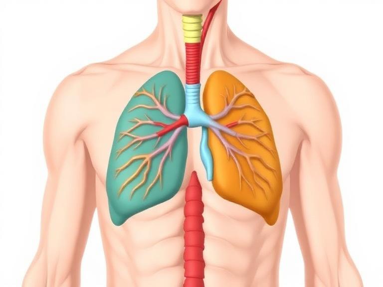
Key Organs Located in the Apothorax
Heart and Major Blood Vessels
The heart sits near the center of the chest cavity, slightly toward the left side. This muscular organ pumps blood through the body’s circulatory system, delivering oxygen and nutrients to tissues.
Major blood vessels connected to the heart include the aorta, pulmonary arteries, and vena cava. These vessels transport blood between the heart, lungs, and the rest of the body.
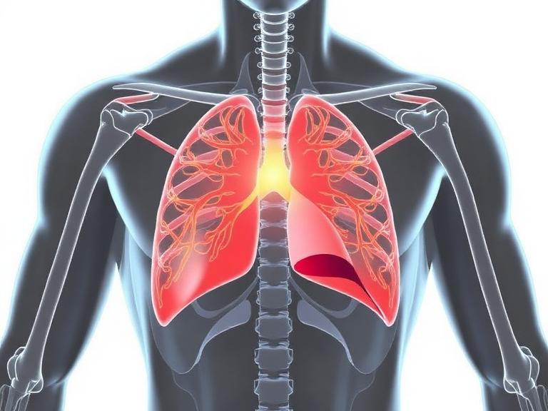
Lungs and Respiratory Structures
The lungs occupy most of the space within the apothorax. Their primary function is to exchange oxygen and carbon dioxide between the air and the bloodstream.
Each lung contains millions of microscopic air sacs called alveoli, where gas exchange occurs. Oxygen passes from these sacs into the bloodstream, while carbon dioxide moves from the blood into the lungs to be exhaled.
Functions of the Apothorax Region
Protection of Vital Organs
One of the primary functions of the apothorax is protecting critical organs such as the heart and lungs. The rib cage acts like a flexible shield, absorbing impacts and preventing damage to the organs inside.
Role in Respiration
Breathing is made possible by coordinated movements within the chest cavity. The diaphragm contracts, the rib cage expands, and the lungs fill with air. This continuous cycle allows oxygen to enter the bloodstream and carbon dioxide to leave the body.
Medical Importance of the Apothorax
Common Diseases and Disorders
Several medical conditions affect the organs located within the apothorax. These include pneumothorax (collapsed lung), pneumonia, pleurisy, and rib fractures.
Doctors often rely on diagnostic tools such as chest X-rays, CT scans, and MRIs to evaluate problems in this region. Early detection of chest diseases can greatly improve treatment outcomes.
Apothorax vs Thorax: Key Differences
| Feature | Apothorax | Thorax |
|---|---|---|
| Definition | Historical term for chest region | Modern anatomical term |
| Usage | Rare in modern medicine | Standard in anatomy and medicine |
| Organs Included | Heart, lungs, blood vessels | Same organs |
| Scientific Acceptance | Mostly historical | Universally recognized |
Conclusion
The apothorax refers to the chest region of the human body—an area responsible for protecting and supporting some of the most vital organs. While modern medicine prefers the term thorax, the concept behind the apothorax remains important for understanding human anatomy. This region houses the heart, lungs, and major blood vessels while enabling essential processes like breathing and blood circulation.
Studying the apothorax reveals how carefully the human body is designed. Bones provide protection, muscles enable movement, and organs work together to sustain life. Even though the terminology has changed over time, the fundamental structure and importance of the chest cavity remain the same.
FAQs
1. What is the apothorax in human anatomy?
The apothorax is an older anatomical term referring to the chest cavity or thorax, which contains vital organs like the heart and lungs.
2. Where is the apothorax located?
It is located between the neck and abdomen, enclosed by the rib cage.
3. What organs are found in the apothorax?
Major organs include the heart, lungs, trachea, esophagus, and major blood vessels.
4. Why is the term apothorax rarely used today?
Modern anatomy replaced it with the standardized term thorax to ensure clear communication in medical science.
5. What separates the apothorax from the abdomen?
The diaphragm, a dome-shaped muscle responsible for breathing, forms the boundary between these regions.