The apothorax refers to the upper region of the human torso commonly known today as the thoracic cavity or chest region. It lies between the neck and the abdomen and contains vital organs that keep the body alive, including the heart, lungs, and major blood vessels. In classical anatomical literature, the word “apothorax” was used to describe the chest area protected by the rib cage and involved in breathing and circulation. Over time, however, modern medical science standardized the term thorax, making “apothorax” a historical or rarely used expression.
Think of the apothorax as the body’s central life chamber. Every breath you take and every heartbeat you feel occurs within this region. When the lungs expand to pull in oxygen, the rib cage widens and the diaphragm moves downward, creating space for air to enter. At the same time, the heart continuously pumps oxygen-rich blood throughout the body. This intricate coordination happens entirely inside the chest cavity. Because these organs are essential for survival, the apothorax is designed with multiple protective structures such as bones, muscles, and connective tissues.
Understanding the apothorax also helps students grasp the bigger picture of human anatomy and physiology. The chest is not just a hollow space; it is a complex structural system that supports breathing, circulation, and protection. Medical professionals examine this region frequently when diagnosing respiratory or cardiac conditions. Even though the term “apothorax” is not widely used today, studying it provides valuable insight into how anatomical terminology has evolved over time.
Historical Origins and Etymology
The word apothorax has roots in ancient Greek language and anatomical terminology. It is derived from two parts: “apo,” meaning away from or upper, and “thorax,” meaning chest or breastplate. Together, these roots describe the upper chest region that supports vital organs and bodily functions. Historically, early anatomists used this term to refer to the central chest cavity before standardized medical terminology was established.
In early medical texts, scholars often used different words to describe the same anatomical structures. Because scientific understanding was still developing, terminology sometimes varied depending on the region, language, or individual researcher. The concept of the apothorax emerged during a time when physicians were trying to explain how breathing and muscular movement worked inside the body. Some early theories even linked the term to processes responsible for energy transfer in muscles and respiratory tissues.
As anatomy evolved into a modern scientific discipline, standardized naming systems were introduced to avoid confusion. Organizations responsible for medical terminology eventually adopted the word thorax as the official name for the chest cavity. This change allowed doctors and researchers worldwide to communicate more clearly about anatomy and disease. Despite this shift, the historical term apothorax still appears in older medical references and educational discussions. Learning about it provides a fascinating glimpse into the history of medical science and how our understanding of the human body has gradually improved.
Location of the Apothorax in the Human Body
Position Between Neck and Abdomen
The apothorax occupies the central section of the upper body, positioned between the cervical region (neck) and the abdominal cavity. This area is enclosed by the rib cage and forms the protective compartment for many of the body’s most critical organs. When you place your hand on your chest and feel your heartbeat or breathing movements, you are touching the outer surface of this region.
From an anatomical perspective, the apothorax acts as a bridge between the upper and lower body systems. Above it lies the neck, where structures such as the trachea and esophagus enter the chest cavity. Below it sits the diaphragm, a dome-shaped muscle that separates the chest from the abdomen. This positioning allows the apothorax to function as the main chamber for breathing and blood circulation.
Another important feature of this region is its structural design. The chest cavity is shaped like a slightly compressed cylinder that widens and narrows during respiration. The intercostal muscles between the ribs expand and contract, enabling the lungs to fill with air. Meanwhile, the diaphragm moves downward to increase the volume of the chest cavity. This dynamic movement makes breathing possible and ensures oxygen reaches every cell in the body.
Because the apothorax lies at the center of multiple body systems, it also serves as a transport corridor. Major blood vessels, nerves, and airways travel through this region. These pathways connect the lungs, heart, digestive organs, and brain, creating a highly coordinated network that keeps the body functioning smoothly.
Boundaries of the Apothorax Region
The apothorax is defined by several anatomical boundaries that help maintain its structure and protect the organs within it. These boundaries include bones, muscles, and connective tissues that form a protective enclosure around the chest cavity.
Superior Boundary
The upper limit of the apothorax is called the thoracic inlet, which connects the chest cavity to the neck. This opening allows important structures such as the trachea, esophagus, nerves, and major blood vessels to pass between the neck and the thoracic cavity. Although it is relatively small compared to the rest of the chest, it plays a crucial role in maintaining communication between different body regions.
Inferior Boundary
The lower boundary of the apothorax is formed by the diaphragm, a large dome-shaped muscle that separates the chest from the abdominal cavity. The diaphragm moves rhythmically during breathing, contracting and flattening as you inhale and relaxing as you exhale. This motion changes the pressure inside the chest cavity, allowing air to flow in and out of the lungs.
Together, these boundaries create a strong yet flexible enclosure for vital organs. The rib cage protects against physical trauma, while the muscles provide mobility for breathing. Without these structural elements, the delicate organs inside the chest would be vulnerable to injury and unable to function efficiently.
Major Structures of the Apothorax
The Rib Cage and Chest Wall
One of the most recognizable structures in the apothorax is the rib cage, which forms the outer framework of the chest. The rib cage consists of twelve pairs of ribs that curve around the chest and attach to the spine at the back. Many of these ribs also connect to the sternum at the front through flexible cartilage, allowing the chest to expand during breathing.
The rib cage serves several essential functions. First and foremost, it acts as a protective shield for the organs inside the chest cavity. The heart and lungs are delicate structures that could easily be damaged without the strong barrier provided by the ribs. Second, the rib cage supports the muscles involved in breathing, including the intercostal muscles that move the ribs up and down.
Another important feature of the chest wall is its ability to adapt to movement. When you inhale deeply, the ribs lift and rotate slightly outward, increasing the volume of the chest cavity. When you exhale, the ribs return to their original position. This movement may seem subtle, but it plays a crucial role in maintaining normal respiration.
Sternum and Thoracic Vertebrae
At the front of the chest lies the sternum, commonly known as the breastbone. This flat bone connects to the ribs and forms the central support of the chest wall. The sternum is divided into three parts: the manubrium, the body, and the xiphoid process. Together, these sections provide stability for the rib cage while protecting the organs behind them.
At the back of the chest are the thoracic vertebrae, which make up the middle portion of the spine. These vertebrae serve as attachment points for the ribs and help maintain the structural integrity of the chest cavity. They also protect the spinal cord, which carries nerve signals between the brain and the rest of the body.
The combination of ribs, sternum, and vertebrae creates a sturdy yet flexible framework. This design allows the chest to withstand physical stress while still permitting the movement required for breathing and other bodily functions.
Organs Located in the Apothorax
The Heart and Circulatory Structures
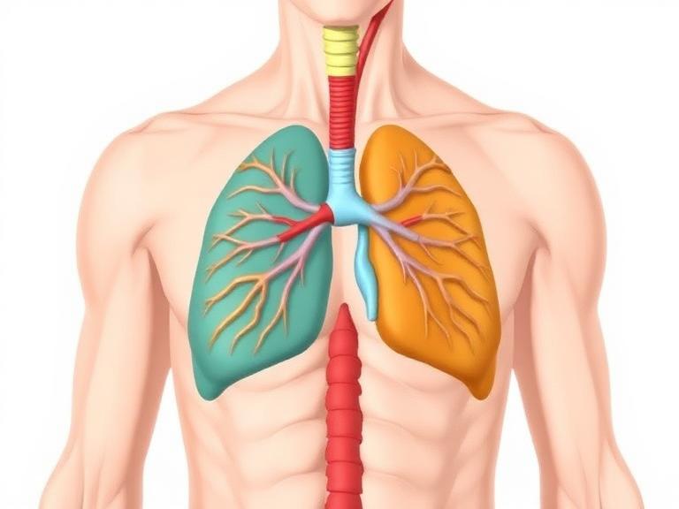
The heart sits at the center of the apothorax, slightly tilted toward the left side of the chest. This muscular organ acts as the body’s main pump, pushing blood through a vast network of arteries and veins. Every minute, the heart beats around 60–100 times in a healthy adult, delivering oxygen and nutrients to tissues throughout the body.
Surrounding the heart are several important blood vessels, including the aorta, pulmonary arteries, and vena cava. These vessels transport blood between the heart, lungs, and other organs. Because of their critical role in circulation, they are carefully protected within the chest cavity.
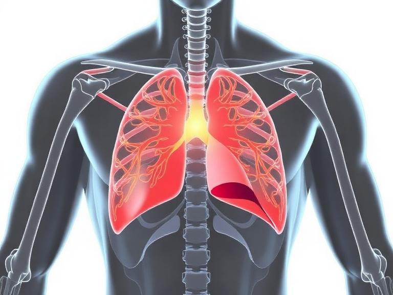
The Lungs and Respiratory System
On either side of the heart lie the lungs, the primary organs of respiration. The lungs are responsible for exchanging oxygen and carbon dioxide between the air and the bloodstream. Each time you inhale, air travels through the trachea and into the lungs, where tiny air sacs called alveoli facilitate gas exchange.
The lungs are surrounded by thin membranes known as pleura, which reduce friction during breathing movements. This design allows the lungs to expand and contract smoothly within the chest cavity.
Functions of the Apothorax
Protection of Vital Organs
One of the most important functions of the apothorax is protecting the organs responsible for breathing and circulation. The rib cage, sternum, and vertebral column form a protective barrier that shields the heart and lungs from injury. Without this structural protection, even minor physical impacts could cause serious damage to these delicate organs.
Role in Breathing and Respiration
Another critical role of the apothorax is enabling respiration. The coordinated movement of the ribs, diaphragm, and chest muscles allows the lungs to expand and contract with each breath. This process ensures that oxygen enters the bloodstream while carbon dioxide is removed from the body.
Apothorax vs Thorax – Understanding the Difference
| Feature | Apothorax | Thorax |
|---|---|---|
| Terminology | Historical anatomical term | Modern standardized term |
| Meaning | Upper chest region | Chest cavity containing vital organs |
| Usage | Rarely used today | Widely used in medicine and biology |
| Organs Included | Heart, lungs, major vessels | Same organs |
The table above highlights how both terms essentially describe the same region of the body. The difference lies mainly in historical usage and scientific standardization.
Why the Term Apothorax Became Obsolete
As anatomical science advanced, medical professionals realized that having multiple names for the same structure could create confusion. To address this issue, global anatomical naming systems standardized terminology and adopted the word thorax as the official term for the chest cavity. This shift ensured that doctors, researchers, and educators around the world could communicate more effectively about the human body.
Clinical Importance of the Apothorax Region
Common Disorders and Medical Conditions
The chest cavity is involved in many medical conditions that affect breathing and circulation. Some of the most common disorders include pneumothorax (collapsed lung), pleurisy, pneumonia, and rib fractures. These conditions can interfere with normal breathing or damage the organs within the chest cavity.
Doctors rely on various diagnostic tools to evaluate problems in this region. Imaging techniques such as chest X-rays, CT scans, and MRIs allow physicians to examine the lungs, heart, and surrounding tissues in detail. Early detection of chest disorders can significantly improve treatment outcomes.
Medical Imaging and Diagnosis in the Apothoracic Area
Modern medicine uses advanced imaging technologies to study the chest cavity and detect abnormalities. Chest X-rays are one of the most common diagnostic tools, allowing doctors to identify lung infections, fractures, or tumors. CT scans provide more detailed images, helping physicians locate problems such as blood clots or structural abnormalities.
Medical imaging plays a crucial role in emergency situations as well. For example, patients experiencing chest pain or difficulty breathing often undergo immediate imaging tests to determine whether the cause is related to the heart, lungs, or surrounding structures.
How Lifestyle Affects the Apothorax and Chest Health
Your daily habits can have a major impact on the health of your chest cavity and the organs within it. Smoking, for instance, damages lung tissue and significantly increases the risk of respiratory diseases. Poor posture can also affect breathing by compressing the chest cavity and limiting lung expansion.
On the positive side, regular exercise strengthens the muscles involved in breathing and improves cardiovascular health. Activities such as swimming, running, and yoga encourage deep breathing, which enhances lung capacity and oxygen delivery throughout the body.
Maintaining a balanced diet, staying physically active, and avoiding harmful habits are some of the most effective ways to protect the organs within the chest cavity. When these habits become part of daily life, the apothorax remains strong and capable of supporting the body’s essential functions.
Conclusion
The apothorax represents the chest region of the human body, a vital anatomical space that houses the heart, lungs, and major blood vessels. Although modern medical terminology now uses the word thorax, the concept of the apothorax still helps us understand the structure and function of the chest cavity. This region serves as both a protective chamber and a dynamic system that enables breathing and circulation.
From the rib cage and sternum to the lungs and heart, every component of the chest cavity works together to sustain life. The complex design of this region demonstrates the remarkable efficiency of the human body. By learning about the apothorax and its role in anatomy, we gain a deeper appreciation for how our bodies function and how important it is to protect these critical systems.
FAQs About Apothorax
1. Is apothorax the same as thorax?
Yes. The term apothorax historically referred to the chest cavity, which is now commonly called the thorax in modern anatomy.
2. Where is the apothorax located in the human body?
It is located between the neck and abdomen, enclosed by the rib cage and separated from the abdomen by the diaphragm.
3. What organs are found in the apothorax?
The chest cavity contains the heart, lungs, major blood vessels, trachea, and esophagus.
4. Why is the term apothorax rarely used today?
Modern anatomy standardized the term thorax to reduce confusion and ensure consistent medical communication.
5. What medical conditions affect the apothorax region?
Common conditions include pneumothorax, pneumonia, pleurisy, rib fractures, and other respiratory or cardiac disorders.